
![]() Contribution of Brain Extracellular Fluid in Mild Damage of Brain Tissue From External Insults
Contribution of Brain Extracellular Fluid in Mild Damage of Brain Tissue From External Insults
PI: Henry W. Haslach, Jr.
The mechanical processes that underlie mild traumatic brain injury from physical impact, shock wave, or sudden acceleration insults are not well understood. Subtle small-scale mechanical damage mechanisms in brain tissue that can modify brain function may be involved in the initial cause of mild traumatic brain injury (mTBI). Another possible contributor, often overlooked in mechanical testing, is the fact that brain tissue is biphasic, composed of water and solid phases, and its mechanical properties are strongly influenced by the high fluid content of the brain tissue itself, whether extracellular or intracellular. The tissue fluid is known to be critical in the mechanical function of other organs.
Brain tissue, which is about 80% water, is composed of roughly equal amounts of neurons and glia, such as astrocytes, (Fig. 1) and a non-load bearing perinueronal net of extracellular matrix proteins that surrounds cellular substructures, but lacks, however, major fibrous structural components. The neurons have protruding dendrites in addition to the signal-conducting axons, whose ends may have several unsheathed branches that communicate with other neurons (Fig 2). Fluid, called the extracellular fluid (ECF), fills the space between these cells; it is this fluid that we postulate transmits the force from external insults to the brain cells.
The pattern of links between neurons, astrocytes and each other forms a mechanically weak network structure that maintains, along with the capillaries, the structural integrity of the brain by a combination of tension in axons, dendrites, and glial processes that is balanced by hydrostatic pressure in the ECF (Van Essen, 1997), rather than any extracellular framework.
The behavior of the ECF that we propose could be one immediate cause of the increase in axonal tension and thus may be a link between the external insult to the brain and cell damage in mTBI.
This project is expected to lead to a mathematical model for the mechanical response of brain tissue and to identify the immediate mechanisms that cause mechanical damage and induce the secondary biochemical effects studied by others. We believe that these results show that a successful mathematical model for brain tissue under insults must account for damage, perhaps through a damage parameter such as permeability, in the range from small to larger strains.
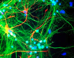
Fig. 1: Rat cortical neurons and glia in mixed tissue culture.
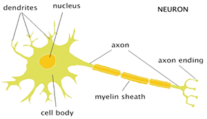
Fig. 2: Neuronal Structure.
Information on the damage mechanisms is needed to interpret the results of any FEM model; knowledge of the location of the maximum stress and strain is not sufficient because a given strain level may cause damage in one brain region and not in another.
