
![]() Damage and Solid-Fluid Interactions during Transient Large Deformation of Rat Brain Tissue in mTBI Study
Damage and Solid-Fluid Interactions during Transient Large Deformation of Rat Brain Tissue in mTBI Study
PI: Dr. Henry Haslach
Since rat models have been routinely used for investigating the initial mechanical causes of mTBI, the characterization of mechanical properties of rat brain tissue must be improved. Brain tissue is composed of water and solid phases so that the mechanical properties of rat brain tissue are strongly influenced by the high water content of the tissue and its flow within the tissue under internal pressure induced by a mechanical load.
Confined compression tests, that enforce uniaxial fluid flow, investigate the uniaxial compressive large deformation mechanical response seen in impact injuries and the solid-fluid interaction within the tissue. The experiments aim to elucidate the role of the intercellular fluid between neurons and glia in mTBI. To deal with the small size of a rat brain and to obtain local information, small specimens are excised, especially focusing on tissue surrounding the hippocampus, which other studies have shown to be particularly susceptible to injury. We have shown that compressive loading, such as due to a shock wave, can damage brain tissue at much lower strains than previously reported. At slow and moderate compressive strain rates, the interstitial fluid carries most of the load until the tissue is sufficiently damaged to permit fluid flow. The constant strain rate stress-strain curves at slow and moderate strain rates reach a stress peak at strains lower than 20%, indicating damage, and the peak magnitude shows a statistically significant dependence on strain rate. An increase in permeability corresponds to softening, and an estimate of permeability suggests it depends on strain rate and as well as on strain. In particular, we hypothesize that intercellular fluid flow in the tissue is the immediate cause of tissue damage by rearranging substructures such as axonal tracts as the applied load opens passageways for fluid transport.
Tests that combine compression and translational shear investigate the tissue large deformation response to the longitudinal and shear components of a pressure wave seen in blast injuries. Translational shear is employed because it is a more likely loading of the brain than rotational shear. The interaction of compression and translational shear involves the intercellular fluid carrying load and therefore applying a local force to the solid component of the tissue that can result in damage.
In constant deformation rate tests up to a 40% translational strain with a normal compressive deformation of 20% of the thickness, no damage from the shear deformation is visible externally, but the translational strain versus stress curves indicate internal damage. The tests at constant strain rates of 0.001, 1, and 100/s show a peak similar to that observed in confined compression. Translational strain at the peak is a statistically significant function of rate but the peak stress magnitude is not. The rate-dependence of the strain at which the peak occurs is more likely a response to the shear and may indicate the strain at which shear damage occurs. Again, the shear damage to the solid component allows the internal fluid to redistribute so that it carries lower load at deformations after the peak is reached.
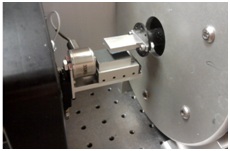
Translational Shear Fixture for 6x6x3 mm Specimens
The Haslach nonlinear viscoelastic (HNV) mathematical model predicts the observed peaks and the coefficients identify appropriate measures of damage. A newly developed unsteady biphasic model, which must also have a damage parameter, is employed to represent the solid-fluid interaction.
